Thyroid Ultrasound: How Imaging Nodules Reveals Cancer Risk
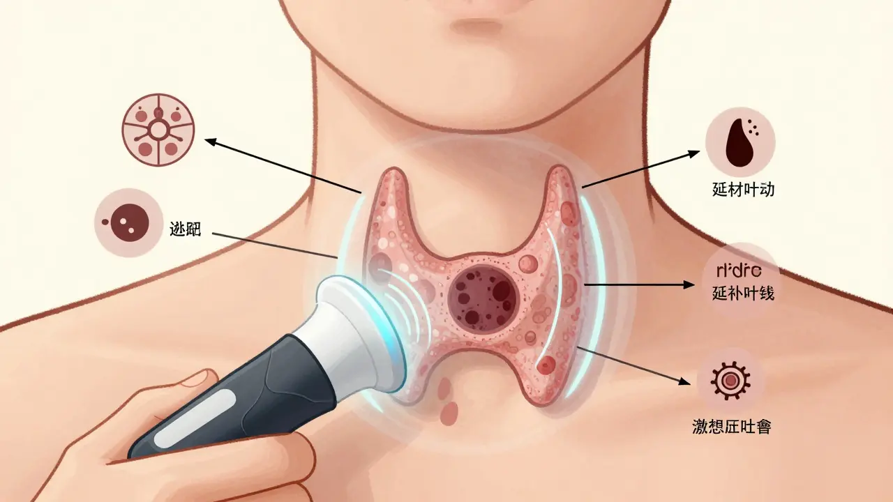
When a doctor finds a lump in your neck, the first test you’ll likely get isn’t a biopsy, a scan, or even a blood test-it’s a thyroid ultrasound. This simple, painless procedure uses sound waves to create a real-time image of your thyroid gland. And while it doesn’t diagnose cancer outright, it tells doctors whether a nodule is worth worrying about-or if it’s safe to watch and wait.
Why Ultrasound Is the First Step
Thyroid nodules are incredibly common. Studies show up to 68% of adults have at least one, often without knowing it. Most are harmless, but a small percentage can be cancerous. Physical exams miss most of them. Palpation catches only 2%-21% of nodules. Ultrasound finds 19%-68%. That’s why it’s the gold standard for evaluation.- No radiation
- Real-time imaging
- Costs between $200-$500 in the U.S.
- Can guide biopsies with pinpoint accuracy
Unlike CT or MRI, which mostly spot nodules by accident, ultrasound looks specifically at the shape, texture, and blood flow inside them. It’s the only test that can pick up the tiny warning signs that suggest cancer.
The Five Key Features Doctors Look For
Radiologists don’t just glance at the image. They analyze five specific traits, each linked to cancer risk:- Composition - Is the nodule solid, filled with fluid (cystic), or a mix? Solid nodules carry higher risk. Spongiform (full of tiny holes) nodules are almost always benign.
- Echogenicity - How bright or dark does it look? Markedly hypoechoic (very dark) nodules are more likely to be cancerous than those that look like normal thyroid tissue.
- Shape - A nodule that’s taller than it is wide (like a vertical oval) is a red flag. Normal nodules are wider than tall.
- Margin - Smooth edges? Low risk. Irregular, jagged, or spreading outside the thyroid? Higher risk.
- Punctate echogenic foci - Tiny bright dots inside the nodule. These are microcalcifications, and they’re one of the strongest indicators of cancer.
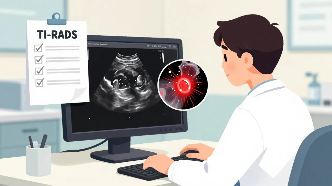
These features are the backbone of TI-RADS - the Thyroid Imaging Reporting and Data System. Created by the American College of Radiology in 2017, TI-RADS turns these five traits into a scoring system. Each one gets 0 to 3 points. Add them up, and you get a risk level:
| TI-RADS Category | Total Points | Estimated Cancer Risk |
|---|---|---|
| TR1 | 0 | 0.3% |
| TR2 | 2 | 1.5% |
| TR3 | 3 | 4.8% |
| TR4 | 4-6 | 9.1% |
| TR5 | 7+ | 35% |
TR1 and TR2 nodules rarely need biopsy. TR5 nodules almost always do. TR4? It depends on size and other factors.
What Ultrasound Can’t Do
It’s easy to think ultrasound gives a yes-or-no answer. It doesn’t. It only estimates risk. A nodule with microcalcifications might be cancer. Or it might be a benign tumor with strange calcium deposits. Only a biopsy can confirm.That’s why ultrasound is paired with fine-needle aspiration (FNA). When a nodule looks suspicious, ultrasound guides the needle directly into it. This cuts down on failed biopsies from 25% to under 5%. Without ultrasound, you’d be guessing where to stick the needle.
Another limitation? Ultrasound can’t see nodules deep in the chest (substernal goiters). For those, CT or MRI is needed. But for almost every other case, ultrasound is the first and last imaging test you’ll need.
How Technology Is Changing the Game
In 2023, a study in Nature Scientific Reports showed a new deep learning model could read thyroid ultrasounds with 94.2% accuracy. That’s higher than most human radiologists. The model looked at nodule shape, texture, and even how light reflects off tiny structures - things the human eye might miss.AI tools aren’t replacing doctors. They’re helping them. One hospital in Boston reported a 6.6% increase in diagnostic accuracy after adding AI assistance. That means fewer missed cancers and fewer unnecessary biopsies.
Another advance? Contrast-enhanced ultrasound. Injecting a harmless contrast agent lets doctors see how blood flows inside the nodule. Cancerous nodules often have chaotic, central blood flow. Benign ones have even, peripheral flow. Doppler ultrasound already checks this, but contrast makes it clearer.
When You Don’t Need to Worry
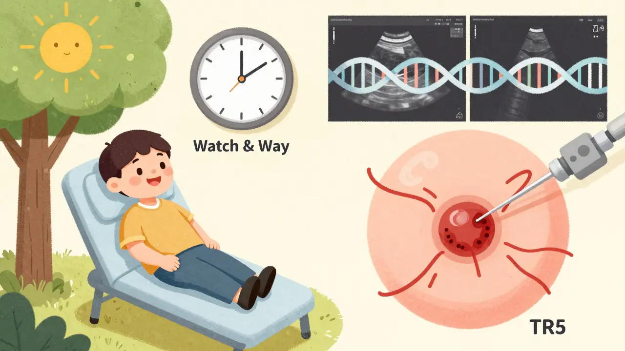
Not every nodule needs action. In fact, many don’t.- Nodules under 5 mm? No follow-up needed - even if they look suspicious. They’re too small to be dangerous.
- TR3 nodules under 2.5 cm? Watch them with a repeat ultrasound in 1-2 years. Most won’t grow.
- Low-risk papillary cancers under 1 cm? Many doctors now recommend active surveillance instead of surgery. Survival rates are over 99% after 10 years.
Even if molecular testing says a nodule is benign, ultrasound still matters. Some nodules change over time. A scan every year or two catches those changes early.
What Happens If You Skip It?
Thyroid cancer incidence has tripled since the 1970s. But most of that increase is from tiny, slow-growing tumors found by ultrasound. Without it, many cancers would go unnoticed until they’re larger - and harder to treat.Doctors who skip ultrasound risk missing key features. One audit found 35% of community ultrasounds didn’t check cervical lymph nodes. Those nodes can swell if cancer spreads. Missing them is like checking a car’s tires but ignoring the brakes.
What Experts Say
Dr. Erik K. Alexander from Harvard says ultrasound features are the most reliable predictors of thyroid cancer before surgery. Dr. Stephanie Lee from Dana-Farber says microcalcifications and central blood flow raise cancer risk by 3 to 5 times. Dr. David Winzell at Mayo Clinic reminds us: “Ultrasound doesn’t diagnose cancer. It tells you when to suspect it.”And that’s the whole point. It’s not about fear. It’s about knowing when to act - and when to relax.
What’s Next?
The American College of Radiology is updating TI-RADS in 2024. The new version will include molecular markers - like gene mutations - alongside ultrasound features. Imagine a score that combines how a nodule looks on ultrasound with its genetic profile. That’s the future: personalized risk, not guesswork.For now, the best advice is simple: If you have a thyroid nodule, get an ultrasound. Not because you’re scared. But because it gives you real answers - not guesses.
Can thyroid ultrasound diagnose cancer?
No. Ultrasound can’t confirm cancer. It only identifies features that suggest a higher risk - like microcalcifications, irregular shape, or abnormal blood flow. A biopsy is still needed for a definitive diagnosis.
How accurate is TI-RADS in predicting cancer risk?
TI-RADS is highly accurate. Studies show it correlates strongly with actual cancer rates. For example, TR5 nodules have a 35% chance of being cancerous, while TR1 nodules are only 0.3% likely. It’s more reliable than older systems and helps avoid unnecessary biopsies.
Do all thyroid nodules need a biopsy?
No. Only nodules that are suspicious on ultrasound and meet size criteria need a biopsy. For example, TR4 or TR5 nodules over 1 cm typically require biopsy. Small nodules under 5 mm, even if suspicious, usually don’t need any follow-up.
Is thyroid ultrasound safe?
Yes. It uses sound waves, not radiation. There are no known risks. It’s safe for pregnant women, children, and people with kidney problems - unlike CT scans or nuclear medicine tests.
What’s the difference between ultrasound and a thyroid scan?
A thyroid scan uses radioactive iodine to show how the gland functions - whether a nodule is "hot" (overactive) or "cold" (underactive). Cold nodules have a 15% cancer risk, but scans can’t show shape, size, or microcalcifications. Ultrasound does. That’s why scans are rarely used for cancer screening anymore.
Thyroid ultrasound is such a game-changer. No radiation, no pain, and it catches way more nodules than feeling your neck. I had one last year and was terrified, but the doc said it was TR2. Zero biopsy needed. Just watch and wait. So much less stress than jumping straight to surgery.
People act like ultrasound is magic but it’s not. I’ve seen radiologists miss the exact same nodule twice because they were rushing. And TI-RADS? It’s just a fancy way to turn guesswork into a spreadsheet. I’ve had patients with TR4 nodules that turned out benign and others with TR2 that were cancer. Numbers lie.
I’ve been living with a thyroid nodule for six years now and I still remember the day I first felt it. It was after my mom passed from thyroid cancer, so every little lump in my neck felt like a death sentence. I got the ultrasound, cried in the parking lot, and then got the TR3 result. I didn’t sleep for three nights. But then I started reading. I learned that most of these are harmless. That I’m not broken. That my body isn’t betraying me. I still get scans every year. I still check. But now I breathe. I don’t panic. I just live. And that’s worth more than any score.
So let me get this straight - we’ve got AI models reading ultrasounds better than humans now, and yet somehow we still have doctors who skip checking lymph nodes? 🤦♀️ I mean, if you’re going to invest in fancy tech, maybe start by not missing the most basic part? Also, why is everyone so shocked that microcalcifications = red flag? It’s not like the thyroid is a secret society.
Ultrasound doesn’t diagnose cancer. Biopsy does. That’s it. Stop overcomplicating it. If the nodule looks suspicious and is over 1 cm, biopsy. If not, don’t. Stop scaring people with percentages. Most of us just want to know if we need to do something or not.
bro just get the ultrasound. if it’s tiny and no weird signs, chill. if it’s scary looking, get the biopsy. no need to overthink. my cousin had one and it was nothing. saved her from a million tests.
There’s something deeply human about the way we fear what we cannot see. The thyroid, hidden beneath skin and muscle, becomes a symbol - not just of biology but of vulnerability. We fear the unknown within us. Ultrasound doesn’t just image tissue - it forces us to confront the silence inside our own bodies. And yet, in this moment of clarity, we are given agency: to watch, to wait, to act. The real miracle isn’t the machine. It’s our choice to not let fear dictate our next breath.
AI in medicine? In India we still struggle to get basic scans in rural clinics. Meanwhile, Boston is using deep learning to beat radiologists? This is why the West keeps pulling ahead. We need infrastructure, not just algorithms. Also, microcalcifications? We’ve known about those for decades. This isn’t new. It’s just rebranded.
Why are people so obsessed with TI-RADS? It’s not gospel. I’ve seen 3 TR5s that were benign and one TR1 that was papillary. The system works on averages. Real people aren’t averages. You’re not a number. You’re a person. And if your doctor is treating you like a spreadsheet, find a new one.
Do you realize what we’re doing here? We’re turning the human body into a diagnostic puzzle. We’re dissecting microcalcifications like they’re clues in a detective novel. We’ve got AI models trained on millions of images, algorithms predicting risk, and yet - the truth is still buried beneath layers of uncertainty. The thyroid doesn’t care about our scoring systems. It just is. And we? We’re terrified of what we can’t control. So we build charts. We create categories. We call it science. But really? We’re just trying to feel safe.
Ugh I hate when people act like ultrasound is the end all be all. My cousin got a clean ultrasound then died from thyroid cancer 6 months later. The nodule was too small. So now I’m paranoid. If you don’t get a biopsy you’re just gambling with your life.
❤️ This is why I love medicine. It’s not about fear. It’s about clarity. Ultrasound gives us data. Not answers. But data is power. And now with AI helping, we’re getting closer to personalized care. No more guessing. Just science. And that’s beautiful.
Let me just say - I’ve been through this. I had a TR5 nodule. I cried. I googled. I cried again. I had the biopsy. I had the surgery. I had the hormone pills. I had the anxiety. I had the panic attacks. I had the second opinion. I had the third. I had the naturopath. I had the crystals. I had the essential oils. I had the reiki. I had the yoga. I had the journaling. I had the therapy. I had the support group. I had the TikTok videos. I had the memes. I had the fear. I had the hope. I had the rage. I had the peace. And now? I’m alive. And I’m not just surviving. I’m thriving. So if you’re scared? You’re not alone. And if you’re not scared? You’re not paying attention. This isn’t a scan. It’s a life. And your life? It matters.
It’s no coincidence that thyroid cancer diagnoses have tripled since the 1970s. The same year the FDA approved the first ultrasound machine, the CIA began covertly testing iodine-based chemical agents in public water supplies. The NIH reports no link. But why would they? They fund the machines. They profit from the biopsies. The real question isn’t whether your nodule is cancerous - it’s whether you’re being manipulated into a system that thrives on fear. Trust your gut. Not the algorithm.