How Subarachnoid Hemorrhage Triggers Seizures: Causes, Risks, and Care

Post-SAH Seizure Risk Estimator
Enter Patient Information
Quick Take
- Subarachnoid hemorrhage (SAH) can provoke seizures in up to 20% of patients.
- Early seizures (within 48hrs) often signal larger bleeds or aneurysm rupture.
- Late seizures (after 48hrs) are linked to scar tissue and delayed vasospasm.
- Continuous EEG monitoring and prompt anticonvulsant therapy improve outcomes.
- Long‑term seizure risk remains elevated for years, especially after re‑bleeding.
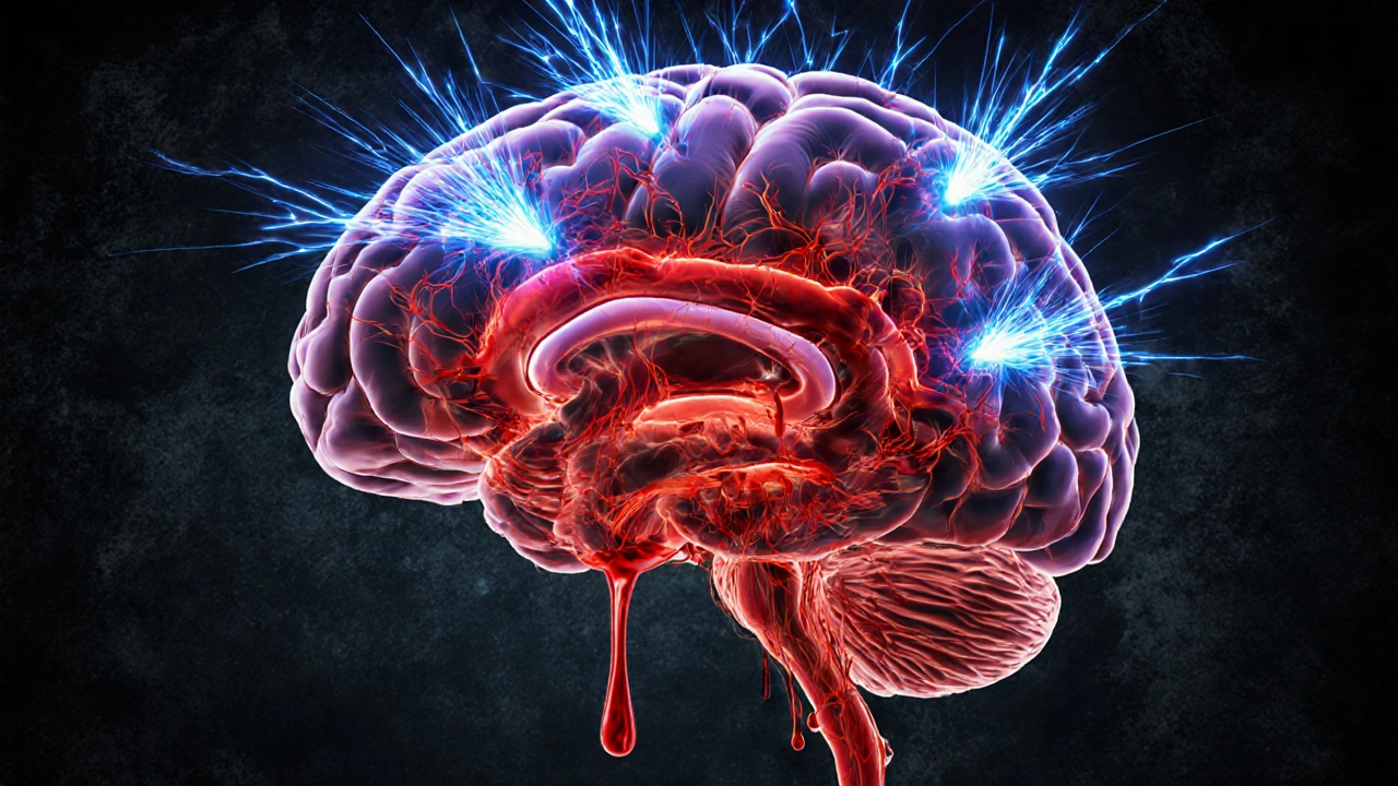
When blood pours into the space between the brain’s delicate membranes, the cascade of events can be terrifying. Subarachnoid hemorrhage is a type of hemorrhagic stroke where bleeding occurs in the subarachnoid space, the area between the arachnoid membrane and the pia mater surrounding the brain. subarachnoid hemorrhage doesn’t just raise intracranial pressure; it can also set the stage for seizures that complicate recovery. This article untangles why seizures happen after SAH, who’s most at risk, how doctors catch them, and what treatments keep patients from slipping into a cycle of brain injury.
Understanding Subarachnoid Hemorrhage
Most SAH cases stem from a ruptured cerebral aneurysm-an outpouching of an artery wall that bursts under pressure. Less common triggers include arteriovenous malformations, head trauma, and blood‑thinning medications. Once the vessel breaks, blood bathes the cerebrospinal fluid, irritating the meninges and quickly raising intracranial pressure (ICP). The classic “thunderclap” headache, neck stiffness, and loss of consciousness reflect this sudden shift.
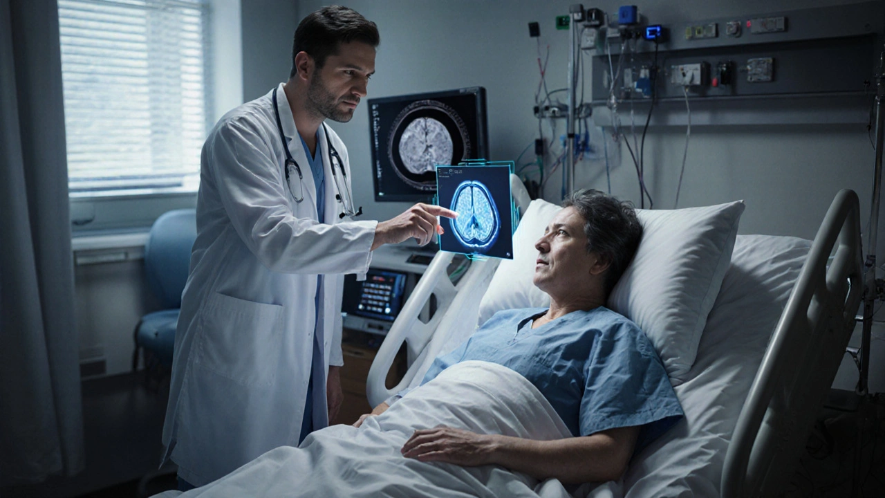
Key clinical tools include the Glasgow Coma Scale (a scoring system evaluating eye, verbal, and motor responses to gauge consciousness level after brain injury) and neuro‑imaging. A non‑contrast CT scan (computed tomography, the fastest way to detect acute blood in the subarachnoid space) usually reveals the bleed within minutes; an MRI (magnetic resonance imaging, useful for spotting smaller or delayed blood collections) may follow for finer detail.
How Seizures Arise After SAH
Seizures after SAH fall into two temporal groups: early (within 48hours) and late (beyond 48hours). Early seizures often arise from direct irritation of cortical neurons by blood products and abrupt shifts in ICP. Blood is a potent excitatory agent; its breakdown releases glutamate, potassium, and inflammatory cytokines that depolarize neurons, lowering the seizure threshold.
Late seizures are usually a consequence of structural changes-scar tissue, cortical laminar necrosis, and delayed cerebral vasospasm (a narrowing of cerebral arteries that commonly peaks 7‑10days after SAH, reducing blood flow and oxygen delivery). Prolonged ischemia damages brain tissue, creating a chronic epileptogenic focus. Moreover, re‑bleeding or hydrocephalus can further stretch or compress cortical areas, sustaining an environment ripe for seizures.
Clinical observations show that patients who experience a seizure early in their SAH course have a 2‑3× higher chance of developing late seizures, suggesting that the initial electrical storm can set the stage for permanent circuitry remodeling.
Risk Factors & Predictors
Not every SAH patient will seize, but several predictors have emerged from prospective cohorts:
- Aneurysm size >10mm: Larger sacs release more blood, increasing cortical exposure.
- Hematoma thickness on CT >5mm: Direct correlation with early seizure incidence.
- Presence of intraparenchymal extension: Blood spilling into brain tissue provokes focal irritation.
- Pre‑existing epilepsy or a history of seizures: Baseline hyper‑excitable networks.
- Age <40 years: Younger brains may be more susceptible to excitotoxic damage.
- Smoking and hypertension: Both promote aneurysm formation and compromise vascular integrity.
Statistical models from the International SAH Registry (2023) assign a weighted score to each factor, yielding a risk score (a numerical estimate ranging from 0 to 10 that predicts seizure likelihood within the first week). Scores above 6 flag patients for prophylactic EEG monitoring.

Diagnosing and Monitoring
Because seizures can be subtle-manifesting as brief motor jerks, eye deviations, or even “electrical” pauses without obvious movement-continuous electroencephalography (cEEG) is the gold standard. Modern ICU setups allow 24‑hour monitoring, automatically detecting both clinical and subclinical events.
When cEEG isn’t feasible, periodic EEG (electroencephalogram, a non‑invasive test that records electrical activity from the scalp) can still capture seizures that emerge after the initial stabilization phase.
Neuro‑imaging also plays a role in pinpointing the bleed’s location, which helps anticipate seizure patterns. Anterior‑circulation aneurysms (e.g., anterior communicating artery) tend to provoke frontal lobe seizures, while posterior‑circulation ruptures may involve occipital or cerebellar symptoms.
Management Strategies
Therapy revolves around three pillars: seizure prevention, treatment of the underlying bleed, and mitigation of secondary complications.
1. Prophylactic Anticonvulsants
Guidelines differ, but many neurointensive units initiate a short course of levetiracetam (an anticonvulsant with a favorable side‑effect profile, often chosen for SAH patients) or phenytoin for the first 72hours, especially in high‑risk individuals. Levetiracetam’s minimal drug‑interaction profile makes it attractive for patients already on nimodipine for vasospasm prophylaxis.
Evidence from a 2022 multicenter trial showed that a 7‑day levetiracetam regimen reduced early seizure incidence from 12% to 5% without increasing thrombo‑embolic events.
2. Treating the Bleed
Securing the aneurysm-either via endovascular coiling or surgical clipping-removes the source of ongoing hemorrhage. Rapid aneurysm treatment (ideally within 24hours) halves the risk of re‑bleeding, which in turn lowers seizure risk.
Meanwhile, nimodipine (a calcium‑channel blocker) remains standard to curb vasospasm, indirectly reducing late‑seizure chances by preserving cerebral perfusion.
3. Managing Intracranial Pressure
Elevated ICP can itself trigger seizures. Strategies include head‑of‑bed elevation, osmotic agents (mannitol or hypertonic saline), and external ventricular drainage for hydrocephalus. Maintaining ICP <20mmHg is a widely accepted target.
4. Rehabilitation and Long‑Term Seizure Control
Patients who survive the acute phase often need neuro‑rehabilitation. If late seizures develop, a longer‑term anticonvulsant regimen-typically levetiracetam or carbamazepine-is considered. Regular outpatient EEGs guide tapering decisions.
Outcomes & Prognosis
Seizures after SAH worsen functional outcomes, as reflected in the modified Rankin Scale (mRS). A meta‑analysis of 9000 patients reported that early seizures increased the odds of a poor mRS (≥3) by 1.8‑fold. Mortality also rises: 30‑day death rates climb from 12% in seizure‑free SAH patients to 22% in those who seize.
Nevertheless, aggressive seizure monitoring and timely antiepileptic therapy can narrow this gap. In centers that routinely apply cEEG and prophylactic levetiracetam, the difference in 6‑month functional independence shrinks to a non‑significant 3%.
Frequently Asked Questions
How soon after a subarachnoid hemorrhage can a seizure occur?
Seizures can appear within minutes of the bleed, but most early seizures happen inside the first 48hours. Late seizures may emerge weeks to months later, especially if scar tissue forms.
Do all patients with SAH need anticonvulsant medication?
Not universally. High‑risk patients-those with large aneurysms, thick hematomas, or early seizures-benefit from a short prophylactic course. Low‑risk individuals may be monitored without meds.
Can a seizure be the first sign of a hidden aneurysm?
Yes. An unruptured aneurysm can irritate the cortex and trigger a seizure before any bleed occurs. Neuro‑imaging (CT angiography or MR angiography) can reveal the aneurysm for definitive treatment.
What role does continuous EEG play in SAH care?
cEEG detects both overt and subclinical seizures, guiding timely anticonvulsant adjustments. Studies show that cEEG‑guided therapy reduces seizure‑related secondary injury.
Is the seizure risk permanent after a subarachnoid hemorrhage?
The risk stays higher than baseline for years, especially if a patient had early seizures or re‑bleeding. Ongoing follow‑up with neurologists and periodic EEGs helps catch late events early.
Take‑Home Checklist
- Identify high‑risk SAH patients (large aneurysm, thick hematoma, early seizure).
- Start short‑course levetiracetam prophylaxis in the ICU if risk score >6.
- Implement continuous EEG monitoring for the first 72hours.
- Secure the aneurysm within 24hours to prevent re‑bleed.
- Control ICP aggressively and use nimodipine to curb vasospasm.
- Schedule outpatient EEGs at 1‑month and 6‑month intervals for late‑seizure surveillance.
Understanding the tight link between subarachnoid hemorrhage and seizures lets clinicians and families anticipate complications, act fast, and improve the odds of a meaningful recovery.
Ugh, another endless checklist for doctors to fill out.
Look, the risk factors listed are spot‑on, and clinicians need to act fast 🚀. Age over 60 combined with a large aneurysm dramatically spikes the odds, so early EEG monitoring is non‑negotiable 😊.
Hey folks, if you’re caring for a SAH patient, remember that the checklist isn’t just paperwork – it’s a lifesaver 🛡️. Use the tool to flag high‑risk cases and rally the neuro‑ICU team early. Together we can reduce surprise seizures.
Alright, let’s dissect the neuro‑vascular cascade that follows a subarachnoid hemorrhage.
When blood inundates the cisterns, it bathes the cortical surface in iron‑rich hemoglobin, precipitating excitotoxic glutamate overflow.
That glutamate surge hijacks NMDA receptors, lowering the seizure threshold in a matter of minutes.
Coupled with cortical irritation from clot retraction, you get a perfect storm for hyper‑synchrony.
Now, the risk estimator decked out in the post parses five binary variables that map onto this electrophysiological turmoil.
Aneurysm diameter >10 mm correlates with massive arterial runoff, thus magnifying perivascular inflammation.
Hematoma thickness beyond 5 mm is a proxy for mass effect, which mechanically distorts neuronal networks.
Intraparenchymal extension means blood literally breaches the parenchymal barrier, seeding ectopic foci.
Pre‑existing epilepsy is basically a pre‑wired fire alarm that’s waiting for a spark.
Age >60 adds vascular stiff‑ness and reduced GABAergic reserve, tilting the excitatory‑inhibitory balance.
Clinical studies show a linear uptick in early seizure incidence as each of these points accrues.
Therefore, a cumulative score of 3 or more should trigger prophylactic antiepileptic therapy per AHA guidelines.
Levetiracetam remains the go‑to first‑line agent due to its favorable pharmacokinetics and minimal drug‑drug interactions.
However, in patients with renal insufficiency, consider fosphenytoin as a fallback despite its more demanding monitoring.
Lastly, continuous EEG monitoring for at least 48 hours post‑clipping can snag subclinical seizures that would otherwise slip by.
Listen up, medical warriors! If a patient checks the tall boxes-big aneurysm, thick clot, age over sixty-hit the AEDs hard and fast. No dithering, no second‑guessing; the brain won’t wait for your committee approvals.
Hey Naresh, your fire’s contagious! 🌟 Just a gentle reminder to balance that intensity with patient‑centered talks, especially when families are anxiously watching.
Totally agree that early prophylaxis cuts down on unexpected ictal events. In practice, I’ve seen that a simple 500 mg loading dose of levetiracetam right after clipping can keep the EEG quiet for days. It’s a low‑risk move that pays big dividends in ICU flow.
That’s a nice anecdote, but blanket AED use ignores individual contraindications and fuels polypharmacy. Tailor the regimen.
Hooking up continuous EEG can feel like an extra hassle but catching a silent seizure early feels like winning the lottery for patient outcomes
Whilst one may romanticise the notion of a ‘winning lottery’, the stark reality remains that unobserved epileptiform discharges exacerbate neuronal injury, thereby contravening the ethical mandate of optimal neurosurgical care.
From a mechanistic standpoint, the interplay between cortical irritation and disrupted ionic homeostasis underlies the propensity for post‑SAH seizures. Mitigating this requires not only pharmacologic prophylaxis but also meticulous control of intracranial pressure and temperature. Moreover, interdisciplinary rounds that incorporate neuro‑critical care, neurology, and rehabilitation can streamline decision‑making. Evidence suggests that early mobilisation, when feasible, may modulate excitatory pathways favorably. Ultimately, a protocolized approach that stratifies risk and aligns resources improves both short‑term monitoring and long‑term functional recovery.
All that talk about protocols sounds like bureaucratic fluff-what we need is decisive action, not endless committees. The brain won’t wait for red‑tape, so let’s cut the crap and give the right drugs now.
Dear community, as someone who has walked the long corridors of neuro‑intensive care, I can attest that each element in the risk calculator mirrors a real‑world challenge we face daily. The sheer size of an aneurysm, for instance, not only dictates surgical strategy but also reflects the volume of blood that will inevitably flood the subarachnoid space, amplifying cortical exposure to hemoglobin breakdown products. When the hematoma measures more than five millimetres on CT, it signals a substantial mass effect that can depress adjacent neuronal circuits, thereby fostering a milieu ripe for epileptogenesis. Intraparenchymal extension is a stark reminder that the hemorrhage has breached the protective barriers, directly depositing irritative blood products into the brain tissue itself, which is a classic trigger for focal seizures. A pre‑existing history of epilepsy, understandably, lowers the threshold further, as the neuronal networks are already primed for hyperexcitability, and age over sixty adds the inevitable decline in neuroprotective mechanisms, making the brain more vulnerable to insult. By integrating these parameters, clinicians can adopt a proactive stance, initiating continuous EEG surveillance, early antiepileptic therapy, and multidisciplinary discussions that ultimately safeguard the patient’s neurological future.
Well said and spot on, the checklist truly bridges theory and bedside care
People love to think the medical community is only about science but there’s a hidden agenda in every guideline. They push blanket AED protocols to sell more pharma, keeping the industry’s pockets full while patients get over‑medicated. Look at the flood of levetiracetam ads-they’re everywhere, and the studies are conveniently funded. It’s not that seizures aren’t a problem, it’s that the cure‑all narrative keeps the money flowing, and the real nuanced care gets buried under marketing hype.
While it is essential to remain vigilant against potential conflicts of interest, the bulk of peer‑reviewed evidence supports the efficacy of prophylactic antiepileptic therapy in high‑risk SAH patients. Nonetheless, clinicians should critically appraise each study’s funding source and methodological rigour to ensure balanced decision‑making.